
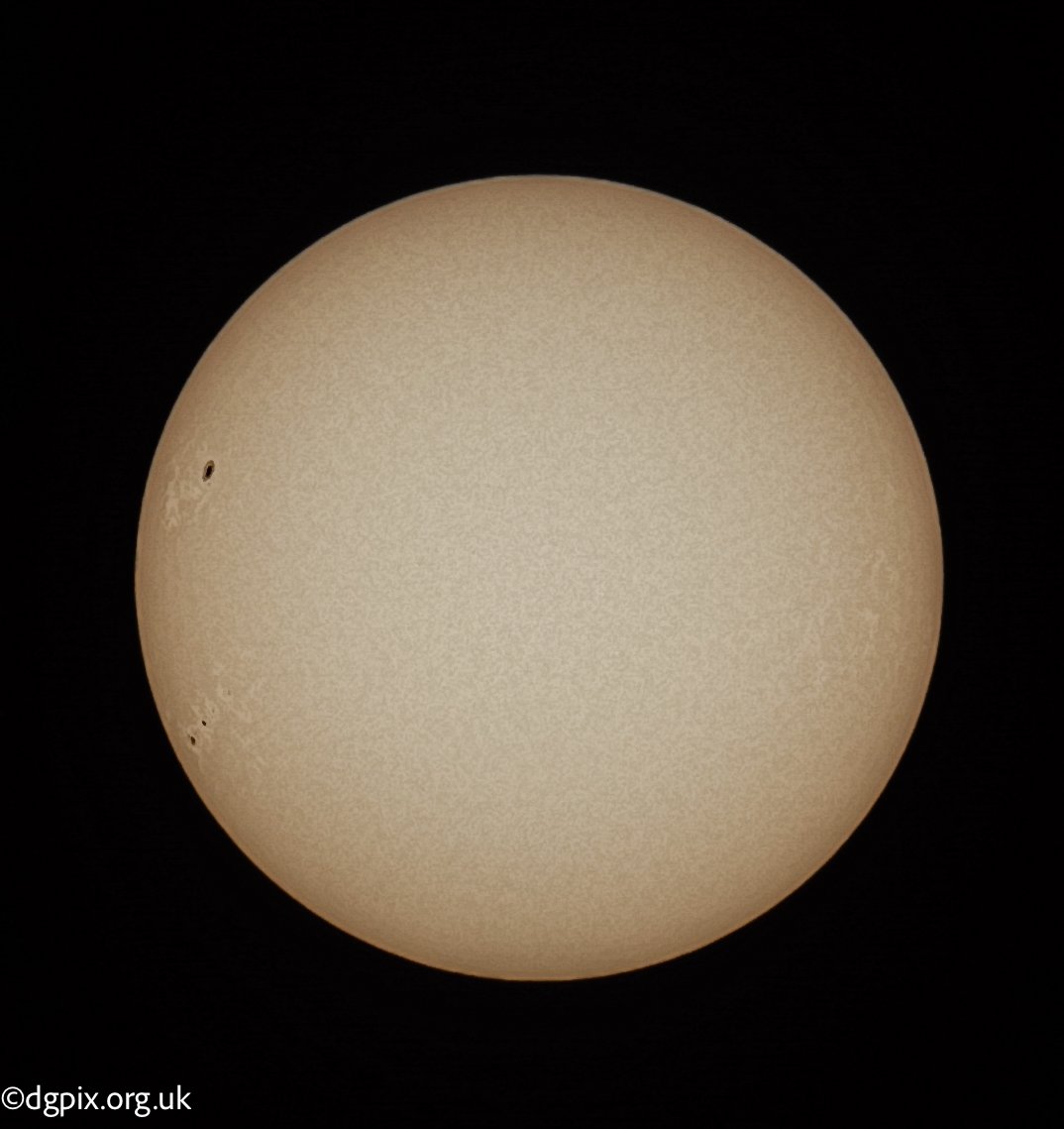
astrophotography
solar
astronomy

In the quiet corners of woodlands and hedgerows, a sinister drama unfolds—one that most passers-by never notice. Flies, seemingly frozen in place on leaves or grass stems, their legs stretched unnaturally, their wings splayed open. These are not ordinary casualties of nature; they are victims of Entomophthora muscae, a fungal parasite that turns its hosts into unwitting puppets before sealing their fate.
Now there are many species of this fungi, and many are determined by the host insect species, but there are some crossovers, and we may have one here. Through observing this particular infected fly in the field and under the microscope, I believe this is E. muscae, even though the host is a dung fly and the species most likely to infect that is E. scatophaga. And of course, as always, I could be wrong! Further reading on this can be found in a paper by Roy et al.,2021
Entomophthora muscae is a pathogenic fungus that specifically targets flies, including species found in Northern Ireland such as commonly found diptera species. The infection begins when fungal spores land on a fly’s exoskeleton. Within hours, the spores germinate, penetrating the insect’s cuticle and spreading through its body.
Once inside, the fungus takes control in a way that seems almost calculated. It invades the fly’s nervous system, altering its behaviour. Infected flies exhibit a phenomenon known as “summit disease,” where they climb to high vantage points—window frames, fence posts, or the undersides of leaves—before succumbing to the infection. Proper "Last Of Us" stuff.....
This particular infection was found in a damp grassland meadow at Brackagh Bog, Co. Armagh.
Yellow Dung fly species showing infection E. m uscae on the abdomen and showing a typical splayed legs and wings pose

Close up macro image of the abdomen and showing
Why do I think this is E. muscae? Well, once this was put under the microscope for a closer look - the conidiophores were visible (the conidia shooters) and a few resting spores were observed as matching the "thick-walled" description in The Fungi of Temperate Europe Vol.2 Laessoe & Peterson, pg 1643. These resting spores are absent in E. scatophaga.
.jpg?width=1463&height=1280&name=0002%20(2).jpg) Microscopy image of the Conidiophore that launches spores
Microscopy image of the Conidiophore that launches spores
.jpg?width=963&height=691&name=0001%20(2).jpg) Close up macro image of the abdomen and showing
Close up macro image of the abdomen and showing
For those interested in documenting this eerie fungal phenomenon, Spring and Summer offer ample opportunities. Observing infected flies in their final moments can provide valuable ecological insights. If you spot a fly with its legs stretched and wings open, consider photographing it and contributing to local biodiversity records, and they also make great subjects for closeup macro photography - they don't fly off!
If you think I am correct or not in this observation, please do get in touch, I'd be more than happy to get more information on this particular type of fungi.
