
Fungi & Slime Moulds Under the Scope
Recording fungi for conservation is essential to understanding their ecological roles, tracking biodiversity changes, and identifying species at risk. Spore morphology, hyphal structures, and reproductive cells often hold the key to distinguishing species. By using a microscope, conservationists can document fungal diversity, contribute to species databases, and detect environmental shifts that impact fungal populations. This detailed recording supports habitat protection efforts and informs broader ecological studies, ensuring fungi receive the recognition they deserve in conservation work.


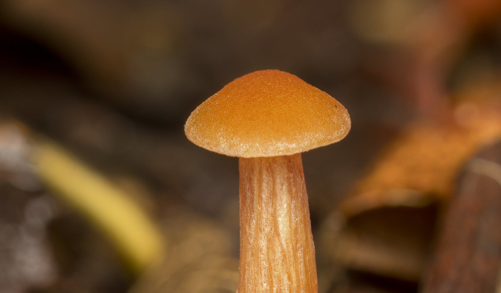
Laccaria laccata
Laccaria laccata - The Deceiver - from a local woodland under the microscope to show identifying features such as spores

Entomophthora muscae
Entomphthora muscae in a yellow dung fly with associated microscopy to show resting spore and conidia
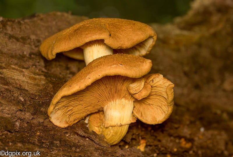
Gymnopilus junonius
Analysis of the Gymnopilus junonius, or Spectacular Rustgill mushroom, a saprobic fungus found in deciduous decaying wood
.jpg?width=2815&height=2215&name=Favolaschia%20calocera%20(7).jpg)
Favolaschia calocera
Discover how this intriguing non-native fungus is spreading in our woods, learn how to identify it, and find out which microscopic features to look for during analysis.

Spinellus fusiger
Discover this mycoparasitic fungi found throughout Northern Ireland. Take a closer look under the microscope to learn how this remarkable fungus functions.

Chlorophyllum rhacodes
The striking Shaggy Parasol, and it's distribution in Northern Ireland and what to look for microscopically when identifying this mushroom